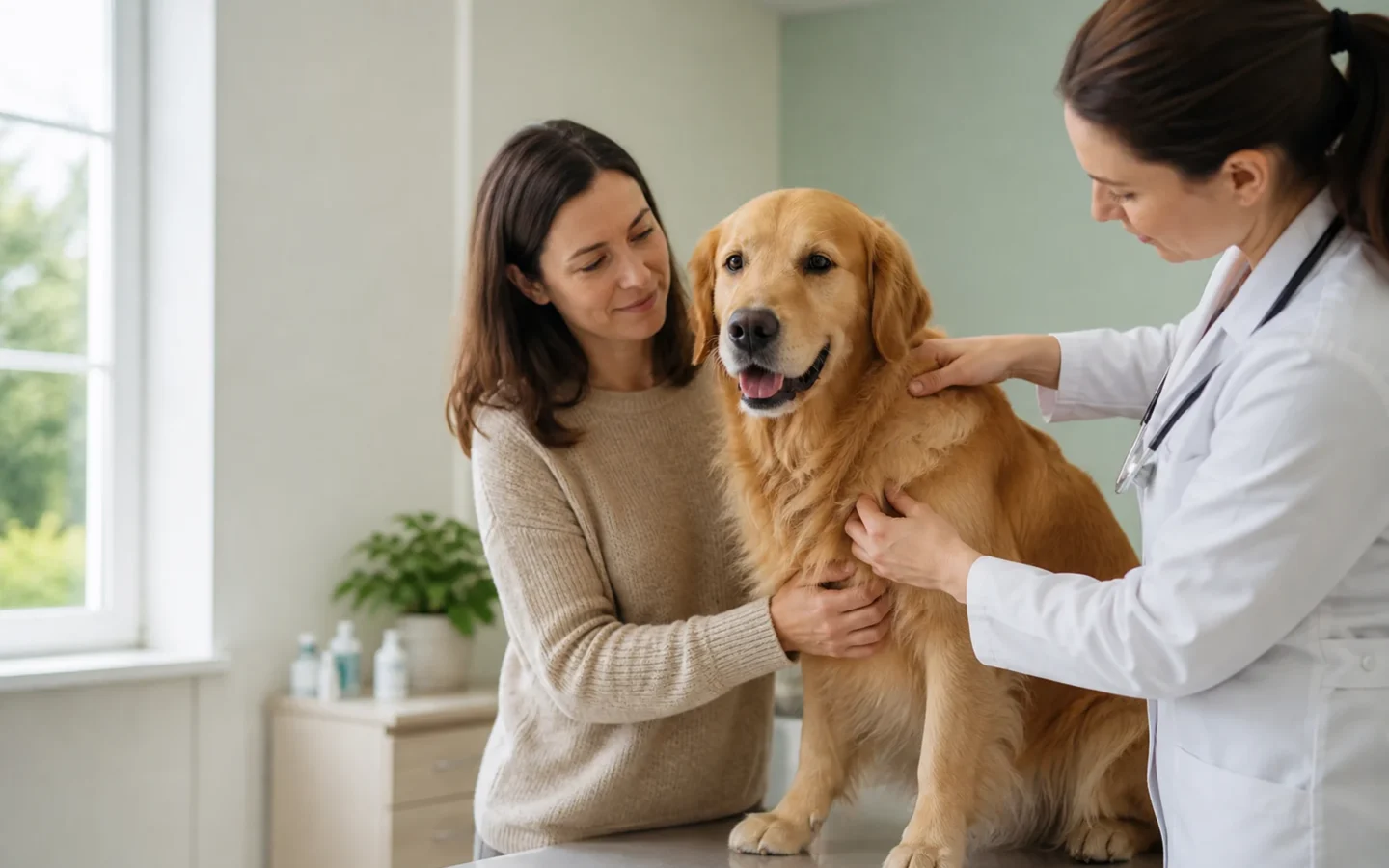
The useful first move: A new lump is a reason to gather good information and arrange a veterinary exam, not a reason to diagnose cancer by touch or by an image search. Many masses are benign; some are inflammatory; others need timely treatment. Microscopic sampling is what begins to separate them.
Finding a lump can make an ordinary evening feel suddenly urgent. Your mind may race ahead to surgery, chemotherapy, or a prognosis before anyone knows what the mass is. A calmer sequence is more useful: document what you can see, protect the area, book the right appointment, and ask what each test will change.
What to do in the first 24 hours
- Take a location photo. Include enough of the body to show exactly where the lump sits.
- Take a close photo with scale. Place a ruler beside, not on top of, the mass. Record length and width in millimeters.
- Write down the texture without squeezing. Is it soft or firm, freely movable or fixed, smooth or irregular, warm, painful, ulcerated, or leaking?
- Check the rest of the dog. Note appetite, energy, breathing, mobility, stool, urination, and recent weight change.
- Protect it. Prevent licking and scratching if the surface is irritated. Do not puncture, drain, massage, or apply human creams.
Those observations create a baseline. They do not tell you whether a mass is benign or malignant. A small mass can be biologically important, while a large soft mass may be a benign lipoma. Growth rate, appearance, and feel help the veterinarian plan the visit, but they cannot replace a cell or tissue diagnosis.
Book the appointment with a useful one-sentence history
When you call, give the clinic a compact summary: “My nine-year-old Labrador has a new two-centimeter firm mass over the right shoulder. I first noticed it three days ago; it is not bleeding, but it seems tender.” Add any whole-body changes and tell the team if your dog takes anticoagulants, steroids, chemotherapy, or other important medication.
Ask how soon the clinic wants to see it and whether the dog should arrive fasted. Do not withhold food, water, insulin, or other medication unless the veterinary team gives specific instructions. Sedation is not always required for sampling, but the plan depends on the mass, the dog, and the procedures that may be considered.
What the veterinarian is trying to learn
The visit usually begins with more than the lump. The veterinarian may examine the skin and mouth, palpate lymph nodes, listen to the heart and lungs, check the abdomen, review weight trends, and look for other masses. This matters because a local-looking problem may sit within a broader health picture, and other conditions can affect sedation, surgery, or treatment choices.
The key questions are:
- Is this a tumor or a non-cancerous process such as inflammation, a cyst, or infection?
- If it is a tumor, what cell type or tissue does it come from?
- How aggressively might it behave locally?
- Is there a reason to look for spread before treatment?
- Will the answer change the surgical approach or the family’s decision?
Fine-needle sampling and biopsy are not the same test
A fine-needle sample collects cells for cytology. It is minimally invasive and often needs no sedation or anesthesia. Cytology can provide a specific answer for some lesions and can categorize others. It may identify inflammation, a lipoma, or certain round-cell tumors, for example. Sometimes the sample contains too few cells or the cell pattern cannot answer every question.
A biopsy collects tissue for histopathology. Tissue architecture can reveal the tumor type with greater confidence and may provide grade, invasiveness, or other prognostic information. Biopsy may mean a small incisional sample, a core, a punch, or removal of the entire mass. The right method depends on location, suspected tumor type, bleeding risk, and the operation that could ultimately be needed.
Why “just take it off” is not always the simplest plan
For some tumors, the first surgery is the best opportunity to remove an appropriate margin of normal-looking tissue around the mass. A narrow, unplanned removal can leave microscopic disease, disturb the surgical field, and make a second operation more difficult. In a tight location such as a limb, face, paw, or body wall, planning matters even more.
Before surgery, ask:
- Would sampling first change how much tissue should be removed?
- If the entire mass is removed, will it be submitted for histopathology?
- Will the laboratory report include surgical margins and grade when relevant?
- What happens if a margin is incomplete?
- Would referral before the first operation preserve more options?
A referral does not commit you to the most intensive treatment. It can simply clarify imaging, biopsy placement, reconstruction, radiation, or what a realistic range of care looks like before an irreversible step.
Staging should answer a decision, not fill a checklist
Staging describes the extent and distribution of cancer. Depending on the diagnosis, it may involve lymph-node sampling, blood and urine tests, chest imaging, abdominal ultrasound, or other targeted tests. Tumor grade and tumor stage are different: grade describes microscopic behavior; stage describes the extent of disease in the patient.
AAHA recommends selecting staging tests for their diagnostic relevance, prognostic value, and fit with the pet’s and family’s needs. A good question is, “If this result is positive or negative, what would we do differently?” That keeps testing connected to anesthesia planning, treatment choice, prognosis, or a meaningful decision.
How to monitor while waiting for the appointment
Do not repeatedly handle the mass. Take a new photo only if it visibly changes, and measure no more than once or twice a week unless the clinic requests otherwise. Record bleeding, discharge, pain, surface breakdown, rapid growth, reduced limb use, appetite loss, or unusual fatigue. A soft recovery collar or clothing barrier may help prevent self-trauma, but it should not compress the mass or overheat the dog.
Do not give ibuprofen, naproxen, acetaminophen, aspirin, leftover antibiotics, or another pet’s medication. Human pain medicines and improvised doses can cause severe toxicity or interfere with planned procedures. Call the veterinary team if comfort is changing.
Read the report in layers
When results arrive, separate four ideas that are easy to blur together:
- Diagnosis: what the cells or tissue are.
- Grade: how the tissue looks microscopically and what that may suggest about behavior.
- Stage: how far disease appears to extend in this dog.
- Margins: whether tumor cells reach the cut edges of a removed specimen.
Ask for a copy of the cytology or pathology report. Request plain-language answers to what is known, what remains uncertain, the likely best and difficult courses, and which next step has the highest chance of changing the outcome. If the findings are complex, a pathology review or oncology consultation may be useful.
This article is educational and cannot diagnose a mass or replace care from a veterinarian who has examined your dog.
开始交流
分享经验或提出有价值的问题;医疗急症请直接联系兽医。